PATHOPHYSIOLOGY IS NOT BACKGROUND KNOWLEDGE FOR THE ADVANCED PRACTICE NURSE. IT IS THE ENGINE THAT DRIVES EVERY CLINICAL DECISION YOU WILL EVER MAKE.
Read that again before you open your textbook or sit down for your first exam.
Because here is what separates the advanced practice nurse who practices safely and excellently from the one who practices mechanically and dangerously — and the difference is not experience, not confidence, and not clinical intuition.
It is the depth of their pathophysiological understanding.
The APN who understands why a patient with heart failure develops pulmonary oedema does not just manage the symptom. They predict which patients are at highest risk, recognise early decompensation before it becomes a crisis, select interventions based on mechanism rather than habit, and explain the treatment rationale to the patient in terms that improve adherence and outcomes.
The APN who understands why a patient with chronic kidney disease is at increased cardiovascular risk does not just order the standard workup. They understand the renin-angiotensin-aldosterone system dysregulation, the anaemia of chronic disease, the mineral and bone disorder, and the accelerated atherosclerosis — and they manage each of those mechanisms simultaneously, proactively, and precisely.
The APN who understands the pathophysiology of sepsis does not wait for the third sign of SIRS to act. They recognise the inflammatory cascade in its early phase, initiate the sepsis bundle, and understand exactly why every element of that bundle targets a specific point in the pathophysiological process.
This is applied pathophysiology at the advanced practice level. It is not memorization of disease descriptions. It is the ability to think mechanistically — to reason from cause to effect, from cellular dysfunction to organ failure, from molecular abnormality to clinical presentation — and to use that reasoning to make better clinical decisions than a nurse without that depth ever could.
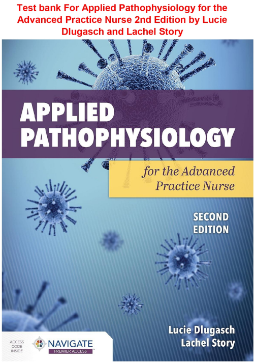
Dlugasch and Story’s Applied Pathophysiology for the Advanced Practice Nurse, 2nd Edition was written for exactly this standard. It is not a medical school pathophysiology text repackaged for nurses. It is a genuinely nursing-focused, APN-level pathophysiology text — built around the clinical decisions advanced practice nurses make, the populations they care for, and the scope of practice they operate within.
This test bank was built to help you master it — and to build the mechanistic clinical reasoning that advanced practice nursing demands.
WHY PATHOPHYSIOLOGY IS THE HARDEST COURSE IN YOUR APN PROGRAM — AND WHY THAT IS THE POINT
Graduate-level pathophysiology is consistently rated as one of the most challenging courses in advanced practice nursing programs. The workload is heavy. The content spans every major organ system. The depth of understanding required goes well beyond what undergraduate physiology and anatomy courses prepared you for.
But here is what nobody says plainly enough: the difficulty is intentional.
Advanced practice nurses make independent clinical decisions. They diagnose. They prescribe. They initiate treatment plans. They manage complex, multi-system patients with multiple comorbidities and overlapping pathophysiological processes. The depth of pathophysiological understanding required to do all of that safely and effectively is enormous — and the difficulty of this course reflects that reality honestly.
The students who understand why the course is hard approach it differently. They do not try to memorize their way through it. They build mechanistic frameworks for each body system — understanding how normal physiology is disrupted, how the body compensates, when compensation fails, how different disease processes interact, and how each of those steps produces the clinical findings they will see in practice.
This test bank is built to accelerate that framework-building process. Every question puts you inside a clinical scenario and asks you to reason from pathophysiological mechanism to clinical decision. Every rationale explains the reasoning step by step. Work through it systematically and the mechanistic thinking that advanced practice pathophysiology is designed to develop will become the way you approach every clinical problem you encounter for the rest of your career.
THE FIVE DIMENSIONS OF APN-LEVEL PATHOPHYSIOLOGICAL REASONING
Undergraduate pathophysiology teaches you what diseases are. Graduate pathophysiology for advanced practice nurses teaches you five things that are far more clinically powerful.
Dimension One — Mechanism at the cellular and molecular level. Why does this happen? Not just that the heart fails — but which mechanisms drive systolic dysfunction versus diastolic dysfunction, and how those different mechanisms produce different clinical presentations and require different management approaches. Not just that inflammation occurs — but which cytokines drive which aspects of the inflammatory response, and how that understanding predicts which patients will progress to septic shock and which will recover.
Dimension Two — Compensation and its limits. Every pathophysiological process triggers compensatory responses. The failing heart activates the RAAS and the sympathetic nervous system. The hypoxic lung attempts to increase respiratory rate and redistribute perfusion. The diabetic kidney increases glomerular filtration pressure to maintain GFR. Understanding compensation means understanding two things simultaneously — why the patient does not yet look as sick as they are, and when compensation will fail and the clinical picture will deteriorate suddenly.
Dimension Three — How disease processes interact. The most complex patients in advanced practice — the ones that require the most sophisticated clinical reasoning — are not the patients with a single diagnosis. They are the patients with five diagnoses that interact in ways that change the presentation, complicate the management, and create risks that exist at the intersection of multiple pathophysiological processes. Diabetes and heart failure. CKD and hypertension and anaemia. COPD and pulmonary hypertension and right heart failure. Pathophysiology at the APN level is the study of intersections, not isolated disease processes.
Dimension Four — From pathophysiology to clinical presentation. The laboratory value, the vital sign, the physical examination finding, the symptom — each of these is the clinical expression of an underlying pathophysiological process. The APN who reads a sodium of 128 and thinks hyponatraemia needs fluid restriction has missed a diagnostic step. The APN who reads a sodium of 128 and thinks about which of the multiple pathophysiological mechanisms that cause hyponatraemia is operative in this specific patient — and then gathers the clinical data that distinguishes them — is practising at the level this course is designed to develop.
Dimension Five — From pathophysiology to clinical decision. Every treatment decision in advanced practice should be mechanistically justified. Every diagnostic test should target a specific pathophysiological question. Every medication choice should reflect an understanding of which mechanism it targets and why that mechanism is operative in this patient. This is applied pathophysiology — the ability to move seamlessly from mechanism to management, from understanding to action, from science to care.
This test bank tests all five dimensions. Not equally in every question — but systematically across the full content of the 2nd edition.
📦 EVERYTHING INSIDE YOUR PURCHASE
Here is precisely what you receive:
- A comprehensive bank of multiple-choice questions covering every unit and chapter of the 2nd edition
- Questions written to reflect the mechanistic clinical reasoning depth of graduate-level advanced practice nursing examinations and APN certification boards
- Every question paired with a clearly identified correct answer
- Detailed rationales explaining the pathophysiological mechanism, the clinical reasoning chain, and the advanced practice decision-making process behind each answer — including specific analysis of why each wrong answer reflects incomplete or inaccurate pathophysiological reasoning
- Questions spanning cellular pathophysiology, genetic and genomic foundations, immune dysfunction, inflammation, every major organ system, and special population considerations in advanced practice
- Both PDF and Word formats included for flexible, multi-device studying
- Content built exclusively around the 2nd edition of Dlugasch and Story — fully updated, evidence-aligned, and APN examination-relevant
📚 COMPLETE CONTENT COVERAGE
Every unit. Every chapter. Every pathophysiological concept and every advanced practice clinical application in the 2nd edition.
Unit 1 — Foundations of Advanced Practice Pathophysiology
- The cell — structure, function, membrane transport, and cellular communication
- Cellular adaptation — hypertrophy, hyperplasia, atrophy, metaplasia, dysplasia
- Cellular injury — reversible and irreversible — causes, mechanisms, and clinical manifestations
- Cell death — necrosis versus apoptosis — pathological versus programmed cell death
- Free radical injury — oxidative stress, antioxidant defences, and clinical implications
- Ischaemia and reperfusion injury — mechanisms and clinical significance in APN practice
- Oedema formation — hydrostatic pressure, oncotic pressure, lymphatic drainage — pathophysiology by type
- Fluid and electrolyte balance — sodium, potassium, calcium, magnesium, phosphate — regulation and dysregulation
- Sodium disorders — hyponatraemia and hypernatraemia — pathophysiology, differential diagnosis, and clinical management
- Potassium disorders — hypokalaemia and hyperkalaemia — mechanisms, ECG changes, and APN management
- Calcium disorders — hypocalcaemia and hypercalcaemia — causes, clinical manifestations, and management
- Magnesium disorders — clinical significance in advanced practice
- Acid-base balance — bicarbonate-carbonic acid system, respiratory and metabolic regulation
- Metabolic acidosis — anion gap and non-anion gap — differential diagnosis using pathophysiological reasoning
- Metabolic alkalosis — causes, compensation, and clinical approach
- Respiratory acidosis and alkalosis — pathophysiology and clinical management
- Mixed acid-base disorders — recognition and APN approach
Unit 2 — Genetic and Genomic Foundations for Advanced Practice
- Basic genetics — DNA structure, gene expression, transcription, and translation
- Types of genetic mutations — point mutations, deletions, insertions, chromosomal abnormalities
- Patterns of inheritance — autosomal dominant, autosomal recessive, X-linked, mitochondrial
- Genetic disorders — single-gene, chromosomal, multifactorial — clinical examples and APN implications
- Epigenetics — gene-environment interactions and clinical significance
- Genomics in advanced practice — pharmacogenomics, genetic risk assessment, cancer genomics
- Genetic testing — types, indications, interpretation, and ethical considerations
- Hereditary cancer syndromes — BRCA1/2, Lynch syndrome, Li-Fraumeni syndrome — APN counselling
- Chromosomal disorders — Down syndrome, Turner syndrome, Klinefelter syndrome — adult health implications
- Teratogenesis — mechanisms, critical periods, and clinical counselling for APNs
- The human microbiome — composition, function, and implications for advanced practice pathophysiology
Unit 3 — Inflammation, Immunity, and Altered Immune Response
- The inflammatory response — innate immunity, acute inflammation, chemical mediators
- Prostaglandins, leukotrienes, cytokines, and complement — mechanisms and clinical significance
- Acute versus chronic inflammation — pathophysiological differences and clinical manifestations
- Wound healing — phases, growth factors, and factors that impair healing
- Chronic inflammation — pathophysiological mechanisms and systemic effects
- The immune system — innate versus adaptive immunity — T cells, B cells, NK cells
- Humoral immunity — B cell activation, antibody classes, and clinical significance
- Cell-mediated immunity — T cell activation, cytotoxic T cells, and immunological memory
- Hypersensitivity reactions — Type I through Type IV — mechanisms, examples, and clinical management
- Type I hypersensitivity — IgE-mediated, anaphylaxis — pathophysiology and emergency management
- Autoimmune disorders — loss of self-tolerance, mechanisms of self-attack, clinical examples
- Immunodeficiency — primary and secondary — pathophysiology and clinical implications for APNs
- HIV pathophysiology — viral entry, CD4 depletion, AIDS-defining conditions, and ART mechanisms
- Systemic lupus erythematosus — immune complex deposition, multi-organ involvement, and clinical management
- Rheumatoid arthritis — autoimmune joint destruction, extra-articular manifestations, and DMARD therapy rationale
- Transplant immunology — rejection mechanisms, immunosuppression rationale, and APN monitoring
- Sepsis and systemic inflammatory response syndrome — pathophysiology, compensated and decompensated shock
- Septic shock — distributive shock mechanism, vasopressor rationale, and Surviving Sepsis Campaign application
Unit 4 — Oncological Pathophysiology
- Cancer biology — hallmarks of cancer, oncogenes, tumour suppressor genes
- Cell cycle dysregulation — CDKs, cyclins, and therapeutic targets
- Tumour suppressor genes — p53, RB, BRCA — mechanisms of loss and clinical significance
- Proto-oncogenes and oncogenes — activation mechanisms and clinical examples
- Carcinogenesis — initiation, promotion, progression — environmental and genetic factors
- Tumour angiogenesis — VEGF pathway and anti-angiogenic therapy rationale
- Metastasis — mechanisms, preferred metastatic sites by tumour type, and clinical implications
- Tumour microenvironment — immune evasion and therapeutic targeting
- Paraneoplastic syndromes — pathophysiology and clinical recognition
- Cancer staging — TNM system and its prognostic significance
- Haematological malignancies — leukaemia, lymphoma, multiple myeloma — pathophysiology and APN management
- Solid tumours — lung, breast, colorectal, prostate, pancreatic — pathophysiology and clinical presentation
- Cancer screening — evidence base, recommendations, and APN role in primary and secondary prevention
- Oncological emergencies — superior vena cava syndrome, spinal cord compression, tumour lysis syndrome, hypercalcaemia of malignancy — pathophysiology and recognition
Unit 5 — Cardiovascular Pathophysiology
Normal Cardiovascular Physiology Review
- Cardiac anatomy and physiology — APN-level review
- Cardiac cycle — systole, diastole, pressure-volume relationships
- Cardiac output determinants — heart rate, preload, afterload, contractility — clinical manipulation
- Coronary circulation — autoregulation and ischaemia mechanisms
- The conduction system — normal and abnormal — pathophysiological basis of dysrhythmias
Atherosclerosis and Coronary Artery Disease
- Atherosclerosis — endothelial dysfunction, lipid accumulation, plaque formation, and vulnerability
- Cardiovascular risk factors — traditional and emerging — pathophysiological mechanisms
- Stable angina versus unstable angina versus acute MI — pathophysiological distinctions
- STEMI — plaque rupture, thrombosis, ischaemia, and infarction — clinical and ECG correlates
- NSTEMI — pathophysiology, biomarker release, and APN management approach
- Complications of MI — arrhythmias, cardiogenic shock, papillary muscle rupture, pericarditis — mechanisms
Heart Failure
- Systolic heart failure — EF-reduced — pathophysiology and neurohormonal activation
- Diastolic heart failure — EF-preserved — pathophysiology and clinical challenges
- Right heart failure — pathophysiology, causes, and clinical manifestations
- Biventricular failure — pathophysiology and clinical presentation
- RAAS activation in heart failure — maladaptive compensation and pharmacological targets
- Sympathetic nervous system in heart failure — beneficial and detrimental effects
- Natriuretic peptides — BNP and NT-proBNP — pathophysiology and clinical utility
- Cardiorenal syndrome — bidirectional pathophysiology and APN management
Hypertension
- Primary hypertension — pathophysiology, genetic and environmental factors
- Secondary hypertension — renal, endocrine, vascular causes — diagnostic approach
- Hypertensive target organ damage — heart, kidneys, brain, retina — mechanisms
- Hypertensive emergency versus urgency — pathophysiological distinction and management
- Resistant hypertension — pathophysiological causes and APN evaluation
Valvular and Structural Disorders
- Aortic stenosis — pressure overload, LV hypertrophy, and clinical deterioration
- Aortic regurgitation — volume overload, LV dilation, and clinical progression
- Mitral stenosis — left atrial hypertension, pulmonary hypertension, and atrial fibrillation
- Mitral regurgitation — volume overload and surgical timing decisions
- Mitral valve prolapse — pathophysiology and clinical significance
Vascular Disorders
- Aortic aneurysm and dissection — pathophysiology, risk stratification, and clinical urgency
- Peripheral arterial disease — atherosclerotic pathophysiology, Ankle-Brachial Index, and clinical management
- Deep vein thrombosis — Virchow’s triad, coagulation cascade, and anticoagulation rationale
- Pulmonary embolism — haemodynamic consequences, right heart strain, and risk stratification
- Pulmonary arterial hypertension — vascular remodelling, pathophysiology, and targeted therapy rationale
Dysrhythmias
- Atrial fibrillation — mechanisms, stroke risk pathophysiology, CHA2DS2-VASc application
- Supraventricular tachycardias — re-entry mechanisms and clinical management
- Ventricular dysrhythmias — VT and VF — pathophysiological substrate and clinical urgency
- Heart block — AV node pathophysiology and clinical implications
- Sudden cardiac death — mechanisms, risk stratification, and primary prevention
Shock
- Haemodynamic framework for shock — preload, afterload, contractility, and oxygen delivery
- Hypovolemic shock — pathophysiology, compensation, and fluid resuscitation rationale
- Cardiogenic shock — pathophysiology, haemodynamic profile, and mechanical support rationale
- Distributive shock — septic, neurogenic, anaphylactic — mechanism-specific management
- Obstructive shock — tamponade and tension pneumothorax — pathophysiology and emergency response
Unit 6 — Respiratory Pathophysiology
Normal Respiratory Physiology Review
- Mechanics of ventilation — compliance, resistance, work of breathing
- Gas exchange — alveolar ventilation, diffusion, V/Q matching
- Oxygen transport — haemoglobin-oxygen dissociation curve and clinical manipulation
- Respiratory control — central and peripheral chemoreceptors and clinical significance
Obstructive Lung Disease
- COPD — emphysema and chronic bronchitis — pathophysiology, hyperinflation, air trapping
- Airflow obstruction mechanisms — mucus hypersecretion, bronchospasm, airway remodelling
- Pulmonary hyperinflation and its cardiovascular consequences
- COPD exacerbation — pathophysiology, antibiotic and corticosteroid rationale
- Asthma — airway inflammation, bronchospasm, airway remodelling — endotypes and phenotypes
- Severe asthma — pathophysiology of near-fatal attack and management
- Bronchiectasis — pathophysiology, recurrent infection cycle, and clinical management
Restrictive Lung Disease
- Intrinsic restrictive disease — interstitial lung disease, idiopathic pulmonary fibrosis — mechanisms
- Extrinsic restrictive disease — pleural effusion, obesity, kyphoscoliosis — mechanisms
- Sarcoidosis — granulomatous inflammation, multi-system involvement, and APN management
Pulmonary Vascular Disease
- Pulmonary embolism — V/Q mismatch, haemodynamic consequences, and right heart strain
- Pulmonary arterial hypertension — vascular remodelling and targeted pharmacotherapy
- Cor pulmonale — right heart failure from pulmonary hypertension
Pleural Disorders
- Pleural effusion — transudative versus exudative — Light’s criteria and pathophysiological differentiation
- Pneumothorax — spontaneous and tension — pathophysiology and clinical urgency
Respiratory Failure
- Type I respiratory failure — hypoxaemic — mechanisms and oxygen therapy rationale
- Type II respiratory failure — hypercapnoeic — mechanisms and ventilatory support rationale
- Acute respiratory distress syndrome — diffuse alveolar damage, inflammatory mediators, and lung-protective ventilation rationale
Lung Cancer
- Small cell versus non-small cell lung cancer — pathophysiology, staging, and targeted therapy
- Paraneoplastic syndromes of lung cancer — SIADH, Cushing syndrome, Eaton-Lambert
Unit 7 — Neurological Pathophysiology
Normal Nervous System Physiology Review
- Neuron structure and function — action potential, synaptic transmission
- Neurotransmitters — glutamate, GABA, dopamine, serotonin, acetylcholine — clinical significance
- Blood-brain barrier — structure, function, and clinical implications
- Cerebral autoregulation — normal and impaired — ICP management implications
- Glial cells — astrocytes, microglia, oligodendrocytes — pathological roles
Cerebrovascular Disease
- Ischaemic stroke — large vessel occlusion, small vessel disease, cardioembolic — pathophysiological differentiation
- Penumbra concept — ischaemic core versus salvageable tissue — thrombolysis and thrombectomy rationale
- Haemorrhagic stroke — intracerebral and subarachnoid — pathophysiology and clinical management
- Cerebral oedema — vasogenic versus cytotoxic — mechanisms and treatment rationale
- Transient ischaemic attack — pathophysiology, ABCD2 score, and secondary prevention
Increased Intracranial Pressure
- Monroe-Kellie doctrine — volume relationships and compensation limits
- Causes and consequences of raised ICP — herniation syndromes and clinical recognition
- Cerebral perfusion pressure — CPP = MAP – ICP — clinical management implications
- Cushing’s triad — pathophysiological mechanism and clinical significance
Neurodegenerative Disorders
- Alzheimer’s disease — amyloid hypothesis, tau pathology, neuroinflammation, and treatment targets
- Parkinson’s disease — dopaminergic depletion, Lewy bodies, and pharmacotherapy rationale
- Multiple sclerosis — demyelination mechanisms, relapsing-remitting versus progressive — disease-modifying therapy rationale
- Amyotrophic lateral sclerosis — upper and lower motor neuron degeneration and clinical progression
- Huntington’s disease — CAG repeat expansion and clinical implications
Seizure Disorders
- Seizure classification — focal versus generalised — pathophysiological basis
- Epileptogenesis — mechanisms of neuronal hyperexcitability and synchronisation
- Status epilepticus — pathophysiology and treatment urgency
- Anticonvulsant mechanisms — sodium channel blockade, GABA enhancement, and others
Traumatic Brain Injury
- Primary versus secondary brain injury — mechanisms and prevention of secondary injury
- Diffuse axonal injury — pathophysiology and clinical outcomes
- Cerebral contusion and haematoma — epidural versus subdural versus intracerebral — mechanisms
Peripheral and Autonomic Nervous System
- Peripheral neuropathy — axonal versus demyelinating — causes and clinical differentiation
- Diabetic neuropathy — pathophysiology and clinical management
- Guillain-Barré syndrome — molecular mimicry, demyelination, and clinical progression
- Autonomic dysfunction — sympathetic and parasympathetic dysregulation — clinical manifestations
Pain Pathophysiology
- Nociception — peripheral sensitisation and central sensitisation
- Neuropathic pain — mechanisms, clinical features, and treatment rationale
- Chronic pain — pathological pain processing and its clinical implications for APNs
Unit 8 — Renal and Urological Pathophysiology
Normal Renal Physiology Review
- Glomerular filtration — GFR, filtration fraction, and autoregulation
- Tubular function — reabsorption, secretion, and concentration mechanisms
- RAAS — renin-angiotensin-aldosterone system — physiological role and pathological activation
- Erythropoietin production — physiological mechanism and anaemia of CKD
- Vitamin D activation — renal role and bone metabolism implications
Glomerular Disease
- Nephrotic syndrome — podocyte injury, proteinuria, hypoalbuminaemia, oedema — mechanism and clinical management
- Nephritic syndrome — haematuria, hypertension, oliguria — pathophysiological distinction from nephrotic
- IgA nephropathy — mesangial IgA deposition and clinical progression
- Membranous nephropathy — anti-PLA2R antibody and clinical significance
- Lupus nephritis — immune complex deposition and WHO classification
Tubular and Interstitial Disease
- Acute tubular injury — ischaemic and nephrotoxic — pathophysiology and clinical differentiation
- Drug-induced nephrotoxicity — mechanisms of aminoglycoside, cisplatin, contrast, and NSAID injury
- Interstitial nephritis — immune-mediated mechanisms and drug causes
- Renal tubular acidosis — types I, II, and IV — pathophysiology and clinical differentiation
Acute Kidney Injury
- AKI staging — KDIGO criteria — clinical application
- Prerenal AKI — volume depletion and reduced renal perfusion — pathophysiology and fluid challenge rationale
- Intrinsic AKI — glomerular, tubular, interstitial, and vascular — pathophysiological differentiation
- Postrenal AKI — obstruction mechanisms and urological decompression rationale
- AKI-to-CKD transition — mechanisms and prevention
Chronic Kidney Disease
- CKD staging — GFR and albuminuria — clinical significance
- Progressive nephron loss — hyperfiltration, proteinuria, and fibrosis — mechanisms
- CKD complications — anaemia, hyperparathyroidism, mineral bone disorder, cardiovascular risk — pathophysiology
- RAAS in CKD — pathological activation and pharmacological targeting
- Hyperkalaemia in CKD — mechanisms, risk factors, and APN management
- Metabolic acidosis in CKD — pathophysiology and treatment rationale
- Dialysis — haemodialysis and peritoneal dialysis — physiological principles
Urological Pathophysiology
- Nephrolithiasis — stone types, formation mechanisms, and metabolic evaluation
- Urinary tract infection — ascending infection, virulence factors, and host defences
- Bladder dysfunction — overactive bladder, underactive bladder — neurological basis
- Benign prostatic hyperplasia — androgen-driven growth, urodynamic consequences
- Urological malignancies — bladder and renal cell carcinoma — pathophysiology and APN recognition
Unit 9 — Endocrine Pathophysiology
Normal Endocrine Physiology Review
- Hormone classes — peptides, steroids, amines — receptor mechanisms and signal transduction
- Hypothalamic-pituitary axis — feedback regulation and APN-level clinical application
Diabetes Mellitus
- Type 1 diabetes — autoimmune beta cell destruction, absolute insulin deficiency
- Type 2 diabetes — insulin resistance, progressive beta cell failure, incretin dysfunction
- Pathophysiology of hyperglycaemia — the ominous octet
- Microvascular complications — retinopathy, nephropathy, neuropathy — AGE mechanism and polyol pathway
- Macrovascular complications — atherosclerosis acceleration and cardiovascular risk
- Diabetic ketoacidosis — insulin deficiency, glucagon excess, ketone production, anion gap acidosis
- Hyperosmolar hyperglycaemic state — extreme dehydration, hyperglycaemia, absence of ketosis — mechanisms
- Hypoglycaemia — counter-regulatory hormone response, hypoglycaemia unawareness — pathophysiology
- Gestational diabetes — placental hormone insulin resistance — maternal and foetal risks
Thyroid Disorders
- Thyroid hormone synthesis, transport, and peripheral conversion — clinical significance
- Primary hypothyroidism — Hashimoto’s thyroiditis — autoimmune mechanism and clinical manifestations
- Hyperthyroidism — Grave’s disease — TSH receptor antibody stimulation and clinical findings
- Thyroid storm — pathophysiology, precipitants, and emergency management
- Thyroid nodules and thyroid cancer — pathophysiology and APN evaluation
Adrenal Disorders
- Cortisol physiology — HPA axis, diurnal variation, and stress response
- Cushing’s syndrome — ACTH-dependent and ACTH-independent — pathophysiology and clinical differentiation
- Primary adrenal insufficiency — Addison’s disease — autoimmune destruction and clinical manifestations
- Addisonian crisis — precipitants, pathophysiology, and emergency management
- Primary hyperaldosteronism — Conn’s syndrome — autonomous aldosterone excess and clinical consequences
- Phaeochromocytoma — catecholamine excess, hypertensive crisis — pathophysiology and clinical recognition
- Congenital adrenal hyperplasia — 21-hydroxylase deficiency — pathophysiology and clinical spectrum
Pituitary Disorders
- Hyperpituitarism — acromegaly, prolactinoma — pathophysiology and clinical manifestations
- Hypopituitarism — causes, hormone deficiency patterns, and clinical management
- Diabetes insipidus — central versus nephrogenic — pathophysiological differentiation
- SIADH — pathophysiology, causes, and clinical management
Metabolic Disorders
- Metabolic syndrome — insulin resistance, visceral adiposity, and cardiovascular risk mechanisms
- Obesity pathophysiology — adipokines, chronic inflammation, and comorbidity mechanisms
- Dyslipidaemia — LDL, HDL, triglycerides — pathophysiological mechanisms and cardiovascular risk
Unit 10 — Gastrointestinal Pathophysiology
Normal GI Physiology Review
- GI motility — enteric nervous system, peristalsis, and segmentation
- Gastric acid secretion — parietal cell physiology, proton pump mechanism
- Digestive enzymes — pancreatic and brush border — clinical significance
- Intestinal absorption — mechanisms and clinical implications of malabsorption
Upper GI Disorders
- GERD — lower oesophageal sphincter dysfunction, acid exposure, and mucosal injury
- Barrett’s oesophagus — metaplasia mechanism, dysplasia progression, and surveillance rationale
- Peptic ulcer disease — H. pylori mechanism, NSAID mechanism, mucosal defence failure
- Gastric cancer — H. pylori carcinogenesis and clinical recognition
Liver Pathophysiology
- Hepatic anatomy and function — detoxification, protein synthesis, bile production
- Acute liver failure — causes, mechanism of hepatic encephalopathy, coagulopathy
- Chronic hepatitis — viral mechanisms — HBV and HCV — liver injury and fibrosis
- Alcoholic liver disease — oxidative stress, acetaldehyde toxicity, Mallory bodies
- Non-alcoholic fatty liver disease and NASH — insulin resistance, lipotoxicity, and fibrosis progression
- Cirrhosis — progressive fibrosis, portal hypertension, hepatic synthetic failure
- Portal hypertension — mechanism, consequences — varices, ascites, hepatic encephalopathy, hepatorenal syndrome
- Hepatocellular carcinoma — cirrhotic and non-cirrhotic pathways, AFP, and clinical recognition
- Drug-induced liver injury — mechanisms, patterns, and clinical significance
Pancreatic Disorders
- Acute pancreatitis — premature trypsinogen activation, autodigestion, systemic inflammatory response
- Chronic pancreatitis — progressive fibrosis, exocrine and endocrine insufficiency
- Pancreatic cancer — KRAS mutation, late presentation, and poor prognosis mechanisms
Lower GI Disorders
- Inflammatory bowel disease — Crohn’s versus ulcerative colitis — pathophysiological differentiation
- Th1 versus Th17 immune mechanisms in IBD — biologic therapy rationale
- Irritable bowel syndrome — gut-brain axis, visceral hypersensitivity, altered motility
- Colon cancer — adenoma-to-carcinoma sequence, mismatch repair deficiency — Lynch syndrome
- Diverticular disease — pathophysiology, diverticulitis mechanism, and clinical management
- Mesenteric ischaemia — arterial and venous — pathophysiology and clinical urgency
Unit 11 — Musculoskeletal Pathophysiology
- Bone physiology — osteoblast and osteoclast coupling, RANK-RANKL-OPG axis
- Osteoporosis — pathophysiology, risk factors, DEXA interpretation, and pharmacotherapy rationale
- Bisphosphonate mechanism — osteoclast apoptosis and clinical application
- Osteoarthritis — cartilage degradation mechanisms, subchondral bone changes, and clinical implications
- Rheumatoid arthritis — synovial inflammation, pannus formation, joint destruction — DMARD and biologic rationale
- Gout — uric acid metabolism, crystal deposition, acute inflammatory response — pharmacotherapy rationale
- Polymyalgia rheumatica and giant cell arteritis — vasculitis mechanisms and corticosteroid rationale
- Ankylosing spondylitis — HLA-B27 association, enthesitis mechanism, and anti-TNF therapy rationale
- Fibromyalgia — central sensitisation, sleep disruption, and clinical management
- Systemic sclerosis — fibrosis mechanisms, vascular involvement, and organ complications
- Paget’s disease of bone — osteoclast hyperactivity, disorganised remodelling, and clinical consequences
- Rhabdomyolysis — muscle fibre breakdown, myoglobinuria, and AKI mechanism
Unit 12 — Haematological Pathophysiology
- Haematopoiesis — stem cell differentiation, growth factors, and clinical manipulation
- Anaemia classification — by mechanism — blood loss, haemolysis, decreased production
- Iron deficiency anaemia — iron absorption physiology, stages of deficiency, and laboratory differentiation
- Anaemia of chronic disease — hepcidin mechanism, iron sequestration, and clinical management
- Megaloblastic anaemia — B12 and folate deficiency — mechanism and neurological consequences of B12 deficiency
- Haemolytic anaemia — intrinsic versus extrinsic — pathophysiological differentiation
- Sickle cell disease — HbS polymerisation, vaso-occlusion, and multi-organ pathophysiology
- Thalassaemia — globin chain imbalance, ineffective erythropoiesis — pathophysiology and clinical management
- Aplastic anaemia — bone marrow failure, autoimmune mechanism, and treatment rationale
- Polycythaemia vera — JAK2 mutation, hyperviscosity, and thrombotic risk
- Coagulation cascade — intrinsic and extrinsic pathways — clinical application in APN practice
- Thrombocytopenia — ITP mechanism, HIT mechanism — pathophysiological differentiation
- DIC — simultaneous thrombosis and haemorrhage — pathophysiology and clinical management
- Venous thromboembolism — inherited and acquired thrombophilias — Virchow’s triad revisited
- Anticoagulation mechanisms — heparin, warfarin, DOACs — pathophysiological rationale for each
Unit 13 — Integumentary Pathophysiology
- Skin anatomy and barrier function — clinical APN significance
- Wound healing — haemostasis, inflammation, proliferation, remodelling — factors affecting each phase
- Pressure injuries — mechanical loading, ischaemia, reperfusion, reactive oxygen species — staging and prevention
- Psoriasis — Th17-mediated epidermal hyperproliferation and biologic therapy rationale
- Atopic dermatitis — barrier dysfunction, Th2 inflammation, and IgE sensitisation
- Melanoma — BRAF mutation, UV carcinogenesis, and targeted therapy rationale
- Non-melanoma skin cancers — actinic keratosis to squamous cell, basal cell mechanisms
- Cellulitis and necrotising fasciitis — bacterial invasion mechanisms and clinical urgency differentiation
- Burns — depth classification, Parkland formula rationale, inhalation injury mechanisms
Unit 14 — Reproductive Pathophysiology
Female Reproductive Pathophysiology
- Menstrual cycle pathophysiology — HPO axis regulation and clinical disruption
- Polycystic ovarian syndrome — insulin resistance, androgen excess, anovulation — mechanism-based management
- Endometriosis — retrograde menstruation hypothesis, peritoneal implantation, and inflammatory mechanism
- Uterine fibroids — oestrogen and progesterone dependence, clinical consequences
- Cervical cancer — HPV oncoproteins E6 and E7, CIN progression — vaccine rationale
- Endometrial cancer — oestrogen-driven type I versus type II mechanisms
- Ovarian cancer — CA-125, BRCA pathways, and late presentation
- Breast cancer — hormone receptor positive, HER2-positive, triple negative — pathophysiological differences and treatment implications
- Menopause — oestrogen deficiency, vasomotor symptoms, cardiovascular and bone consequences
- Osteoporosis in menopause — accelerated bone loss mechanism and HRT rationale
Male Reproductive Pathophysiology
- Benign prostatic hyperplasia — androgen-driven, stromal and glandular growth — alpha-blocker and 5-ARI rationale
- Prostate cancer — androgen signalling, Gleason grading, PSA kinetics, and androgen deprivation mechanism
- Erectile dysfunction — vascular, neurological, and endocrine mechanisms — PDE5 inhibitor rationale
- Testicular pathophysiology — varicocoele, torsion, and cancer — APN recognition
Sexually Transmitted Infections
- HIV — viral life cycle, CD4 tropism, ART mechanism classes
- HPV — oncogenic types, E6/E7 oncoproteins, cancer progression
- Chlamydia and gonorrhoea — intracellular and extracellular mechanisms, PID pathophysiology
- Syphilis — Treponema pathophysiology, stages, and neurological complications
- Herpes simplex virus — latency and reactivation mechanisms, neonatal infection risk
Unit 15 — Special Populations and Advanced Practice Considerations
- Paediatric pathophysiology differences — developmental physiology and its clinical significance for APNs
- Geriatric pathophysiology — physiological ageing, frailty, and multi-system interactions
- Immunosenescence — clinical implications for infection risk and vaccine response
- Sarcopaenia — mechanisms and clinical consequences in older adults
- Pregnancy pathophysiology — maternal adaptations, placental physiology, and obstetric complications
- Preeclampsia — placental dysfunction, VEGF imbalance, endothelial injury — pathophysiology and management
- Gestational diabetes — pathophysiology and maternal-foetal consequences
- COVID-19 and post-COVID pathophysiology — ACE2 receptor entry, multi-organ involvement, long COVID mechanisms
- Environmental and occupational pathophysiology — toxin exposure, carcinogen mechanisms, and APN counselling
- Obesity as a pathophysiological driver — adipokine excess, chronic inflammation, and comorbidity mechanisms
🎯 WHO THIS TEST BANK IS FOR
Graduate nursing students in NP, DNP, or CRNA programmes who are enrolled in a graduate-level pathophysiology course and want comprehensive, mechanistic exam practice aligned to Dlugasch and Story’s 2nd edition.
Students using Applied Pathophysiology for the Advanced Practice Nurse as their assigned textbook who want questions designed to match the clinical reasoning depth, APN focus, and content sequencing of this specific edition.
Advanced practice nursing students preparing for NP certification examinations — AANP FNP-C, ANCC FNP-BC, ANCC AGPCNP-BC, ANCC AGACNP-BC — who want focused pathophysiology practice at the level of mechanistic clinical reasoning these examinations require.
CRNA students who want to consolidate their pathophysiological understanding of the organ systems most critical to anaesthesia practice — cardiovascular, respiratory, renal, and neurological — at the APN level.
PhD and DNP students in advanced coursework who want to review and deepen their pathophysiological foundation before clinical practicums or comprehensive examinations.
Advanced practice nurses in clinical practice who want to refresh and deepen their pathophysiological knowledge base systematically — by organ system, by clinical population, or by disease mechanism.
Faculty teaching graduate-level pathophysiology who need a comprehensive, APN-level question bank for building course examinations, clinical reasoning assessments, and comprehensive evaluations.
💡 BUILDING THE MECHANISTIC REASONING THAT ADVANCED PRACTICE DEMANDS
There is a cognitive shift that the best APN students make early in their graduate pathophysiology course — and the ones who make it earliest consistently outperform their peers on examinations and, more importantly, in clinical practice.
The shift is from descriptive thinking to mechanistic thinking.
Descriptive thinking says: heart failure causes fluid retention, dyspnoea, and peripheral oedema. Mechanistic thinking says: reduced cardiac output activates the RAAS because the kidneys sense reduced perfusion pressure, aldosterone increases sodium and water reabsorption, the resulting fluid overload raises pulmonary capillary wedge pressure, fluid transudates into the alveoli producing dyspnoea, and the increased hydrostatic pressure in peripheral capillaries drives fluid into the interstitium producing oedema.
Both statements describe the same clinical reality. But only one of them equips you to understand why ACE inhibitors and mineralocorticoid antagonists improve survival in heart failure, why loop diuretics reduce preload and alleviate pulmonary oedema, why beta-blockers paradoxically improve outcomes despite reducing cardiac output acutely, and why a patient with heart failure who develops worsening renal function on aggressive diuresis may actually be experiencing cardiorenal syndrome rather than diuretic-induced dehydration.
Mechanistic thinking does not just describe clinical presentations. It predicts them. It explains treatment rationale. It anticipates complications. It distinguishes patients who look similar on the surface but have fundamentally different underlying processes.
This test bank is built to develop mechanistic thinking — not to reward descriptive recall. Every question starts with a clinical scenario. Every rationale traces the reasoning from mechanism to clinical finding to management decision. Every wrong answer is analysed to show which step in the mechanistic chain it gets wrong.
Work through it with a mechanistic lens and this course will permanently change how you think about every patient you see.
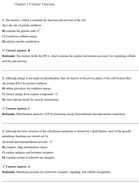
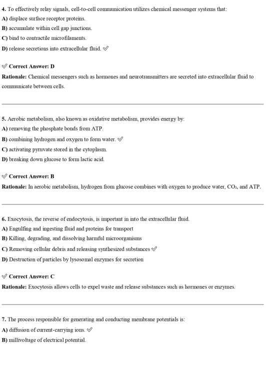
📝 10 SAMPLE QUESTIONS
These are real questions from the full test bank. They reflect the graduate-level pathophysiological depth, clinical reasoning focus, and APN application orientation of the complete product.
Question 1
An NP is evaluating a 67-year-old patient with a long history of poorly controlled hypertension who presents with a blood pressure of 220/130 mmHg, altered mental status, and bilateral papilloedema on fundoscopic examination. The patient’s creatinine has risen from a baseline of 1.2 to 2.8 mg/dL over the past 24 hours and troponin is mildly elevated. Which pathophysiological mechanism best explains the multi-organ involvement in this patient?
- A. Chronic atherosclerotic damage causing simultaneous deterioration across multiple vascular beds
- B. Hypertensive emergency-induced fibrinoid necrosis of arteriolar walls causing end-organ ischaemia in the brain, kidneys, and heart simultaneously
- C. Acute decompensation of pre-existing heart failure causing reduced perfusion to all organs
- D. Autoimmune vasculitis triggered by chronic hypertension causing multi-organ involvement
Correct Answer: B Rationale: Hypertensive emergency is defined by severely elevated blood pressure — typically above 180/120 mmHg — with acute target organ damage. The pathophysiological mechanism is fibrinoid necrosis of arteriolar walls — a process in which extremely elevated intravascular pressure overwhelms autoregulatory capacity, disrupts endothelial integrity, and triggers a cascade of platelet aggregation, fibrin deposition, and arteriolar wall destruction. This fibrinoid necrosis causes ischaemia in the end-organs supplied by those arterioles simultaneously — the brain producing hypertensive encephalopathy and cerebral oedema evidenced by altered mental status, the kidneys producing acute kidney injury evidenced by the rising creatinine, and the myocardium producing demand ischaemia evidenced by the troponin elevation. The bilateral papilloedema confirms raised intracranial pressure from cerebral oedema. Chronic atherosclerosis causes gradual deterioration rather than acute simultaneous multi-organ failure. Heart failure is not the primary process here — it may be a consequence. Vasculitis is not the mechanism in hypertensive emergency.
Question 2
An FNP is evaluating arterial blood gases from a patient with a three-day history of vomiting due to gastric outlet obstruction. The results are: pH 7.51, PaCO2 49 mmHg, HCO3 38 mEq/L, serum potassium 2.9 mEq/L. Which pathophysiological explanation best accounts for all four abnormal values?
- A. Respiratory alkalosis from hyperventilation with metabolic compensation causing hypokalaemia
- B. Metabolic alkalosis from HCl loss in vomit, renal HCO3 retention, hypokalaemia-driven renal H+ secretion, and respiratory hypoventilation as compensation
- C. Mixed metabolic alkalosis and respiratory acidosis from sedative use during prolonged vomiting
- D. Metabolic alkalosis from contraction of the extracellular fluid volume without renal compensation
Correct Answer: B Rationale: This is a complex acid-base scenario that requires mechanistic reasoning across multiple physiological systems. The elevated pH confirms alkalosis. The elevated HCO3 identifies the primary process as metabolic alkalosis. The elevated PaCO2 confirms appropriate respiratory compensation — hypoventilation retains CO2 to buffer the alkalosis. The metabolic alkalosis has three concurrent pathophysiological mechanisms in this patient. First, vomiting causes loss of HCl — hydrochloric acid — depleting hydrogen ions from the body and raising pH. Second, the volume depletion from vomiting activates the RAAS, increasing aldosterone and driving renal sodium retention with concurrent hydrogen and potassium secretion — perpetuating the alkalosis and the hypokalaemia simultaneously. Third, hypokalaemia itself drives hydrogen ions into cells in exchange for potassium to maintain electroneutrality, reducing extracellular hydrogen ion concentration and further alkalising the blood while also stimulating renal hydrogen secretion over potassium secretion — a phenomenon called paradoxical aciduria. This interconnected mechanism is the hallmark of contraction alkalosis sustained by hypokalaemia.
Question 3
An AGACNP is managing a 72-year-old patient with decompensated heart failure. The patient’s BNP is 1,840 pg/mL, blood pressure is 98/64 mmHg, heart rate is 112 bpm, and urine output has declined to 18 mL per hour over the last three hours despite IV furosemide. The creatinine has risen from 1.4 to 2.1 mg/dL. Which pathophysiological process best explains the worsening renal function in the context of aggressive diuresis?
- A. Furosemide nephrotoxicity from prolonged exposure causing direct tubular injury
- B. Cardiorenal syndrome type 1 — acute haemodynamic compromise from the failing heart reducing renal perfusion pressure, compounded by diuresis-induced prerenal azotaemia and elevated renal venous pressure from right heart failure
- C. Contrast nephropathy from a prior imaging study causing delayed AKI
- D. Obstructive uropathy from prostate enlargement causing postrenal AKI during aggressive diuresis
Correct Answer: B Rationale: Cardiorenal syndrome type 1 describes acute worsening of renal function occurring in the setting of acute cardiac decompensation. In this patient, the pathophysiology is multimechanistic. The severely reduced cardiac output reduces renal perfusion pressure — a prerenal mechanism. However, the unique haemodynamic feature of heart failure — particularly with right heart dysfunction — is elevated central venous pressure transmitted backward to the renal veins. Elevated renal venous pressure reduces the transrenal perfusion gradient — the difference between renal arterial pressure and renal venous pressure — more profoundly than reduced arterial pressure alone. Aggressive diuresis reduces preload and may further reduce cardiac output in a patient already on the flat portion of the Starling curve, paradoxically worsening renal perfusion. The interaction of reduced forward flow, elevated venous back-pressure, and neurohormonal vasoconstriction from RAAS and sympathetic activation creates a haemodynamic environment where the kidneys receive inadequate net perfusion despite — and partly because of — the diuretic therapy. Furosemide nephrotoxicity does not occur with short-term therapeutic use. Contrast nephropathy and obstructive uropathy are not supported by the clinical scenario.
Question 4
A family NP is seeing a 54-year-old patient with a recent diagnosis of primary hyperaldosteronism — Conn’s syndrome. The patient has hypertension resistant to three antihypertensive medications and a serum potassium of 2.8 mEq/L despite oral replacement. Which sequence of pathophysiological events best explains the clinical presentation?
- A. Adrenal cortex tumour produces excess cortisol causing sodium retention, hypertension, and hypokalaemia through glucocorticoid receptor stimulation
- B. Autonomous aldosterone excess from adrenal adenoma stimulates renal principal cells to upregulate luminal sodium channels and basolateral sodium-potassium ATPase, causing sodium and water retention raising blood pressure, and driving potassium and hydrogen secretion into the tubular lumen causing hypokalaemia and metabolic alkalosis
- C. Excess aldosterone increases angiotensin II levels which directly constrict peripheral vasculature causing hypertension independent of sodium retention
- D. Elevated aldosterone suppresses renin causing reduced angiotensin II and secondary vasoconstriction that raises blood pressure while promoting renal potassium wasting through direct tubular toxicity
Correct Answer: B Rationale: Primary hyperaldosteronism — most commonly caused by a unilateral adrenal adenoma — produces autonomous aldosterone excess independent of the normal RAAS regulatory feedback. Aldosterone acts on the principal cells of the late distal tubule and cortical collecting duct by binding to the mineralocorticoid receptor, which translocates to the nucleus and upregulates the transcription of ENaC — the epithelial sodium channel — on the luminal membrane and the sodium-potassium ATPase on the basolateral membrane. The result is increased sodium reabsorption, which drives water reabsorption and expands extracellular fluid volume — raising blood pressure. The electrochemical gradient created by sodium reabsorption drives potassium and hydrogen secretion into the tubular lumen through ROMK channels and hydrogen-ATPase pumps respectively — producing hypokalaemia and metabolic alkalosis. The autonomous nature of aldosterone production means renin is suppressed — not elevated — differentiating primary from secondary hyperaldosteronism. Cortisol excess causes Cushing’s syndrome, not Conn’s syndrome. Aldosterone does not directly increase angiotensin II — it is the product of RAAS activation, not its upstream driver.
Question 5
An NP is caring for a 44-year-old patient with type 1 diabetes who presents with fruity breath, Kussmaul respirations, blood glucose of 480 mg/dL, pH 7.19, anion gap of 26 mEq/L, and serum bicarbonate of 9 mEq/L. What is the pathophysiological sequence that produced the Kussmaul respiratory pattern?
- A. Hypoxaemia from pulmonary oedema triggered by diabetic cardiomyopathy stimulating tachypnoea
- B. Absolute insulin deficiency causing unrestrained lipolysis, free fatty acid oxidation to acetyl-CoA, ketone body accumulation, anion gap metabolic acidosis, and peripheral chemoreceptor stimulation driving deep rapid ventilation to eliminate CO2 and partially compensate the acidosis
- C. Hyperglycaemia directly stimulating the respiratory centre in the brainstem causing hyperventilation
- D. Severe dehydration from osmotic diuresis reducing cerebral perfusion and triggering a stress respiratory response
Correct Answer: B Rationale: Kussmaul respirations — deep, rapid, laboured breathing — in DKA represent the respiratory compensation for severe metabolic acidosis. The pathophysiological chain begins with absolute insulin deficiency in type 1 diabetes. Without insulin, glucagon is unopposed — lipolysis accelerates in adipose tissue, flooding the liver with free fatty acids. Hepatic beta-oxidation of these free fatty acids generates excess acetyl-CoA, which cannot enter the TCA cycle efficiently and is instead shunted toward ketogenesis — producing acetoacetate, beta-hydroxybutyrate, and acetone. Ketone bodies are organic acids that dissociate at physiological pH, releasing hydrogen ions that consume bicarbonate — reducing serum bicarbonate and producing anion gap metabolic acidosis. The falling pH — in this case 7.19 — stimulates peripheral chemoreceptors, which activate the respiratory centre to increase ventilatory rate and depth. This removes CO2 from the blood, raising pH toward normal — partial compensation. Kussmaul breathing is therefore not a primary respiratory disorder but a maximal compensatory respiratory response to metabolic acidosis. The fruity breath is from volatile acetone — a ketone product — being exhaled. Hyperglycaemia alone, dehydration, and cardiac dysfunction do not produce Kussmaul respirations through the mechanisms described in the wrong options.
Question 6
An AGPCNP is evaluating a 78-year-old patient with a three-week history of progressive confusion, worsening headache, and gait instability. The patient has no history of trauma but takes warfarin for atrial fibrillation. CT head shows a hypodense crescent-shaped lesion compressing the right hemisphere. Which pathophysiological mechanism best explains this lesion and its slow clinical onset?
- A. Ischaemic stroke from an embolic event causing cortical infarction with oedema in a crescent distribution
- B. Chronic subdural haematoma — bridging vein rupture from minor or unrecognised trauma, slow venous bleeding into the subdural space, initial clot liquefaction and osmotic expansion, worsening mass effect over weeks — potentiated by anticoagulation
- C. Epidural haematoma from middle meningeal artery laceration with the classic lucid interval and rapid neurological deterioration
- D. Hypertensive intracerebral haemorrhage causing deep white matter haemorrhage with surrounding oedema
Correct Answer: B Rationale: The clinical picture — insidious onset over weeks, progressive cognitive decline, headache, and gait disturbance in an older anticoagulated patient — combined with the CT finding of a hypodense crescent-shaped lesion is the classic presentation of chronic subdural haematoma. The pathophysiological mechanism begins with rupture of bridging veins — thin-walled venous structures that traverse the subdural space from the brain surface to the dural sinuses. In older adults, cerebral atrophy stretches these bridging veins across a greater distance, making them more susceptible to rupture from minor or even unrecognised trauma. The initial haematoma clots but then undergoes fibrinolysis over days to weeks, liquefying into a protein-rich hygroma. The osmotic gradient created by the breakdown products draws cerebrospinal fluid and plasma into the subdural space, causing the collection to expand — worsening mass effect and neurological symptoms over time. Warfarin anticoagulation both facilitates the initial bleed and impairs haematoma resolution. The hypodense appearance on CT reflects the chronic, liquefied nature of the collection. Epidural haematoma involves arterial bleeding, rapid expansion, and the classic lucid interval — not a three-week subacute progression.
Question 7
An FNP is evaluating a 51-year-old woman with a six-month history of fatigue, cold intolerance, constipation, weight gain of 8 kg despite no dietary change, and a TSH of 48 mIU/L with a free T4 of 0.4 ng/dL. The patient has positive anti-TPO antibodies. Which pathophysiological mechanism best explains this presentation?
- A. Secondary hypothyroidism from pituitary TSH deficiency causing thyroid gland atrophy and reduced T4 production
- B. Primary autoimmune thyroiditis — Hashimoto’s disease — in which anti-TPO and anti-thyroglobulin antibodies target thyroid peroxidase and thyroglobulin, lymphocytic infiltration destroys follicular cells, T4 production falls, TSH rises through loss of negative feedback, and the clinical syndrome of hypothyroidism develops
- C. Thyroid hormone resistance syndrome in which normal T4 and T3 levels are present but peripheral receptor insensitivity causes clinical hypothyroidism with elevated TSH
- D. Iodine deficiency causing reduced thyroid hormone synthesis, compensatory TSH elevation, and goitre formation as the primary mechanism
Correct Answer: B Rationale: The clinical presentation — fatigue, cold intolerance, constipation, weight gain, markedly elevated TSH, suppressed free T4, and positive anti-TPO antibodies — is the textbook presentation of Hashimoto’s autoimmune thyroiditis, the most common cause of primary hypothyroidism in iodine-sufficient regions. The pathophysiological mechanism involves molecular mimicry or loss of central tolerance to thyroid antigens — particularly thyroid peroxidase, an enzyme critical to thyroid hormone synthesis. Anti-TPO antibodies and cytotoxic T lymphocytes infiltrate the thyroid gland, causing lymphocytic thyroiditis and progressive destruction of thyroid follicular cells. As follicular cell mass decreases, T4 production falls. The fall in circulating T4 removes negative feedback on the hypothalamic-pituitary axis, driving TSH progressively higher in an attempt to stimulate residual thyroid tissue. The resulting TSH of 48 mIU/L with suppressed free T4 confirms primary — not central — hypothyroidism. Secondary hypothyroidism would show low TSH, not elevated. Thyroid hormone resistance is rare and would not show the antibody profile described. Iodine deficiency is uncommon in regions with iodised salt.
Question 8
An AGACNP is managing a 62-year-old patient with non-ST-elevation myocardial infarction who develops sudden haemodynamic collapse on day two of hospitalisation. Echocardiography shows a new, large ventricular septal defect with left-to-right shunting and biventricular dysfunction. Which pathophysiological mechanism best explains this catastrophic complication?
- A. Reperfusion injury from primary PCI causing myocardial stunning and acute heart failure without mechanical complication
- B. Infarct expansion and myocardial wall rupture — coagulative necrosis of the interventricular septum at the infarct zone reaching full-thickness necrosis by day two to five, with mechanical disruption creating an abnormal communication between the left and right ventricles — causing sudden left-to-right shunting, right ventricular volume overload, and biventricular failure
- C. Acute mitral regurgitation from papillary muscle dysfunction causing acute pulmonary oedema without septal involvement
- D. Pericardial tamponade from free wall rupture causing haemodynamic collapse with equalisation of diastolic pressures
Correct Answer: B Rationale: Ventricular septal rupture is one of the most catastrophic mechanical complications of acute MI, typically occurring two to five days after the index event — the period when coagulative necrosis is maximal and myocardial integrity is at its lowest before scar formation begins. The pathophysiological sequence involves full-thickness necrosis of the interventricular septum at the infarct zone. Without the structural integrity of viable myocardium, the high left ventricular systolic pressure — typically 120 mmHg — compared to the right ventricular systolic pressure — typically 25 mmHg — creates a mechanical shear force that ruptures the necrotic septum. The resulting abnormal communication creates massive left-to-right shunting — volume overloading the right ventricle — causing right ventricular failure, pulmonary overcirculation, reduced systemic cardiac output, and biventricular haemodynamic collapse. Echocardiographic colour Doppler showing left-to-right septal flow confirms the diagnosis. Reperfusion injury and myocardial stunning do not produce an anatomical VSD. Papillary muscle rupture causes acute MR — a different echo finding. Free wall rupture causes tamponade — not a septal defect with left-to-right shunting.
Question 9
An NP is evaluating a 38-year-old male patient with a three-day history of progressive ascending weakness beginning in the lower extremities, loss of deep tendon reflexes, and mild facial weakness. The patient had a Campylobacter jejuni gastroenteritis four weeks ago. CSF shows elevated protein with normal cell count — albumino-cytological dissociation. What is the pathophysiological mechanism responsible for this presentation?
- A. Direct Campylobacter invasion of peripheral nerves causing bacterial axonitis and ascending motor paralysis
- B. Post-infectious molecular mimicry in which antibodies generated against Campylobacter lipooligosaccharides cross-react with ganglioside epitopes — specifically GM1 — on peripheral nerve myelin or axons, causing immune-mediated demyelination or axonal injury of peripheral motor and sensory nerves — Guillain-Barré syndrome
- C. Campylobacter toxin-mediated destruction of anterior horn cells causing lower motor neuron paralysis resembling poliomyelitis
- D. Vitamin B12 deficiency from malabsorption during the gastroenteritis causing subacute combined degeneration of the spinal cord
Correct Answer: B Rationale: The clinical presentation — progressive ascending flaccid paralysis, areflexia, and cranial nerve involvement two to six weeks after a febrile gastrointestinal illness, with CSF showing elevated protein but normal cell count — is classic Guillain-Barré syndrome. The pathophysiological mechanism is post-infectious molecular mimicry. Campylobacter jejuni is the most common infectious trigger for GBS, accounting for approximately 30 to 40 percent of cases. The outer membrane lipooligosaccharides of Campylobacter share structural homology with gangliosides present on peripheral nerve membranes — particularly GM1, GD1a, and GalNAc-GD1a. The immune response generated to clear the infection produces antibodies that cross-react with these ganglioside epitopes on peripheral nerves, triggering complement-mediated attack and Fc-receptor-dependent immune cell recruitment. The axonal variant — AMAN — targets axonal gangliosides directly, while the demyelinating variant — AIDP — targets Schwann cell membrane components. The albumino-cytological dissociation — high protein from blood-nerve barrier disruption and protein leak, normal cell count because this is an antibody-mediated rather than inflammatory cell-mediated process — is a hallmark diagnostic finding. Direct bacterial invasion, toxin-mediated anterior horn injury, and B12 deficiency do not produce this specific clinical and CSF profile.
Question 10
An FNP is evaluating a 47-year-old patient with HIV who is adherent to ART with an undetectable viral load and a CD4 count of 420 cells/mcL. The patient’s annual screening reveals a new microalbuminuria with urine albumin-to-creatinine ratio of 58 mg/g and eGFR of 71 mL/min/1.73m2. The patient has well-controlled hypertension on lisinopril and no diabetes. Which pathophysiological mechanism most likely explains the renal findings?
- A. HIV-associated nephropathy — collapsing focal segmental glomerulosclerosis — from direct HIV viral protein injury to podocytes despite ART suppression
- B. Tenofovir disoproxil fumarate nephrotoxicity — mitochondrial dysfunction in proximal tubular cells causing Fanconi syndrome with tubular proteinuria, phosphate wasting, and declining GFR — distinguished from glomerular proteinuria by the pattern of renal involvement
- C. Lisinopril-induced acute interstitial nephritis causing glomerular filtration decline and proteinuria
- D. Age-related nephrosclerosis causing a gradual decline in GFR that is coincidental to the HIV diagnosis
Correct Answer: B Rationale: In the ART era, HIV-associated nephropathy — the classic collapsing FSGS from direct viral podocyte injury — has become less common in virologically suppressed patients, while ART-related nephrotoxicity has emerged as an important cause of renal dysfunction. Tenofovir disoproxil fumarate — TDF — is nephrotoxic through a specific mechanism. TDF accumulates in proximal tubular cells where it inhibits mitochondrial DNA polymerase gamma, impairing mitochondrial replication and causing mitochondrial dysfunction. This impairs the high-energy demands of proximal tubular reabsorption, resulting in Fanconi syndrome — a generalised proximal tubular defect characterised by phosphaturia, glucosuria at normal blood glucose, aminoaciduria, and low-molecular-weight proteinuria. Progressive injury reduces GFR. The proteinuria in TDF nephrotoxicity is tubular in origin — not glomerular — which can be distinguished by urine protein electrophoresis. Tenofovir alafenamide — TAF — is a newer formulation with lower intracellular TDF levels and significantly less nephrotoxicity. The APN managing HIV patients must monitor for this complication with regular eGFR and urine albumin-to-creatinine ratio screening and consider ART regimen modification when nephrotoxicity is suspected.
4 STUDY STRATEGIES FOR GRADUATE-LEVEL PATHOPHYSIOLOGY MASTERY
The Mechanism Chain Method
For every pathophysiological process you encounter in this test bank — every disease, every complication, every clinical syndrome — build a mechanism chain before you answer the question. Start at the cellular or molecular level. Follow the chain through tissue dysfunction, organ dysfunction, systemic compensation, and clinical manifestation. For heart failure: reduced contractility → reduced cardiac output → reduced renal perfusion → RAAS activation → sodium and water retention → increased preload → pulmonary oedema → dyspnoea. For every disease. Every time. This is slower at first. It becomes automatic. And when it does, you will find that you can reason through clinical scenarios you have never seen before — because you are following the chain of physiological logic rather than retrieving a memorised answer.
The Compensation Failure Pivot
One of the most clinically important and consistently tested concepts in graduate pathophysiology is the transition from compensated to decompensated disease. For every major chronic condition in this test bank — heart failure, CKD, COPD, cirrhosis — identify the three most important compensatory mechanisms the body uses, then identify the clinical findings that signal each mechanism is failing. This pivot point is where patients deteriorate acutely, where hospitalisation occurs, and where APN clinical decisions have the highest stakes. Understanding it deeply is the difference between recognising early decompensation and reacting to a crisis.
The Differential Mechanism Framework
Advanced practice examinations frequently present clinical scenarios in which two or more diagnoses share superficially similar presentations but differ in underlying mechanism — and that mechanistic difference determines the correct management. Nephrotic versus nephritic syndrome. Prerenal versus intrinsic AKI. Respiratory versus metabolic acidosis with compensation. Cardiac tamponade versus tension pneumothorax. For each of these pairs, build a side-by-side comparison based on mechanism rather than symptom checklist. Then apply it to every relevant question in this test bank. Over time this comparative mechanistic thinking becomes the basis of your differential diagnosis reasoning in clinical practice.
The Treatment Rationale Reverse Engineering Protocol
One of the most powerful ways to deepen pathophysiological understanding is to work backward from treatment to mechanism. Pick any pharmacological treatment in this test bank’s clinical scenarios — an ACE inhibitor in heart failure, spironolactone in primary hyperaldosteronism, dexamethasone in cerebral oedema, N-acetylcysteine in TDF nephrotoxicity. Ask yourself: why does this treatment work? Which specific step in the pathophysiological chain does it target? What would happen if you gave this treatment to a patient with a different but superficially similar condition? This reverse engineering process builds the kind of mechanistic pharmacological understanding that graduate-level pathophysiology is designed to develop — and that APN practice demands.
❓ FREQUENTLY ASKED QUESTIONS
Is this the official Jones & Bartlett Learning publisher test bank for the 2nd edition? No. This is an independently developed study resource. It is not affiliated with Jones & Bartlett Learning or the authors of Applied Pathophysiology for the Advanced Practice Nurse. It is a supplementary study product designed to support students and educators using the 2nd edition.
My graduate programme uses a different pathophysiology textbook. Will this test bank still be useful? The mechanistic pathophysiological content covered in this test bank is consistent with the core content of all major graduate-level APN pathophysiology curricula. The questions are built around the organ system organisation and clinical application focus of Dlugasch and Story’s 2nd edition, but the underlying pathophysiological mechanisms are universal. Students using other APN pathophysiology texts will find the vast majority of content directly applicable.
What formats are included with my purchase? Both PDF and Word formats are included. PDF is ideal for reading on any device without formatting disruption. Word allows you to select questions by organ system or chapter, adapt clinical scenarios for your patient population, and build course assessments if you are a faculty member.
How quickly will I receive my file after purchasing? Your download link is sent to your email automatically the moment your payment is confirmed. No manual processing is required. Most students have their file open within minutes of completing checkout.
Is this test bank useful for NP certification examination preparation? Yes — and specifically so. The AANP FNP-C and ANCC FNP-BC certification examinations test pathophysiological understanding as the foundation for clinical assessment, differential diagnosis, and management decisions across all major organ systems. The mechanistic clinical reasoning that this test bank builds is precisely what those examinations test at the advanced practice level. Use it as part of a comprehensive certification preparation plan that also includes clinical management review and full-length practice examinations.
Can I use this test bank if I am in a CRNA or DNP programme? Yes. CRNA students will find the cardiovascular, respiratory, renal, and neurological pathophysiology content particularly valuable for understanding the physiological consequences of anaesthesia and surgery. DNP students will find the organ system coverage and population-based pathophysiology content relevant across their programme and for comprehensive examination preparation.
I am a nursing faculty member teaching graduate pathophysiology. Can I use this for course examinations? Absolutely. All questions are written at graduate level with detailed, mechanistic rationales. The Word format makes it straightforward to select questions by organ system, adapt clinical scenarios for your specific patient population, and organise questions into structured examinations. Many graduate faculty in advanced practice nursing programmes use independently developed test banks to supplement their examination question pools — particularly for clinical application and mechanistic reasoning content.
What if something is wrong with my file or my order? Contact our support team directly with your order details and a description of the issue. We respond promptly and resolve every problem without delay. Your access to this resource should be seamless from the moment of purchase.
🏁 THE LAST WORD — FOR THE ADVANCED PRACTICE NURSE IN TRAINING
You chose advanced practice nursing because you wanted to go deeper.
Deeper into the clinical science. Deeper into the diagnostic reasoning. Deeper into the patient relationship. Deeper into the responsibility of independent clinical decision-making. Deeper into the possibility of making a difference at a level that requires mastery, not just competence.
Applied pathophysiology is the course that builds the foundation that depth requires. It is the hardest course in your programme for a reason — because the clinical authority you are preparing for demands a level of mechanistic understanding that cannot be faked, abbreviated, or bypassed.
Dlugasch and Story wrote their textbook for the APN who takes that responsibility seriously. They did not write a survey of disease descriptions. They wrote a clinically applied, mechanistically rigorous preparation for the kind of practice that changes patient outcomes.
This test bank was built in that spirit. Every question is a clinical encounter. Every rationale is a teaching moment. Every wrong answer is an opportunity to identify and correct a gap in mechanistic reasoning before that gap appears in clinical practice.
Use it rigorously. Use it consistently. Let it change how you think — not just how you answer questions.
The patients who will sit across from you in your practice deserve an advanced practice nurse who prepared with that level of seriousness.
You are becoming that nurse.

Josesa8065 –
Extremely helpful study resource
Dennys –
Great value for money
Rosemary637 –
Made revision much easier
Zippysouthgate –
Clear, concise, effective
Duncan N. –
Made a huge difference in my studies
bryan kim –
I feel very lucky to have come across this gem
Winnie M. –
Perfect!
Grace W. –
what a gem
Lucille Maria –
I like it
Gerald N –
Very very helpful
Norah Helen –
An excellent study resource
Helen Christie –
Really helped me pass my finals
Caleb Martins –
A very helpful resource
Dorcas M. –
I can now face my finals with confidence
Liam Mbogo –
Excellent quality content.
Lisa Horton –
Made studying much easier
Terence V. –
Explanations helped me understand every mistake
Even Njeri –
Comprehensive coverage of key nursing subjects.
Bruno Molino –
Helped me identify areas needing more review.
Mary Lincoln –
A practical resource for focused studying
Mary Kesh –
An excellent test bank, very helpful for pathophysiology exam preparation.
Alicia Silva –
Great questions that improved my nursing knowledge and exam confidence.
Bruno Molino –
Accurate practice questions with useful explanations.