The human body is the most extraordinary system ever assembled.
Twelve organ systems working in seamless coordination. Trillions of cells communicating through electrical signals and chemical messengers. A cardiovascular system that pumps blood through 60,000 miles of vessels. A nervous system that processes sensory input, generates motor commands, stores memories, and creates consciousness — all simultaneously. A renal system that filters 180 liters of fluid every day, returning 99% of it to the circulation while precisely regulating the composition of every drop.
Understanding this system — at the molecular level, the cellular level, the tissue level, the organ level, and the integrated systems level simultaneously — is not optional for healthcare professionals. It is the foundation upon which every clinical decision, every patient assessment, and every therapeutic intervention is built.
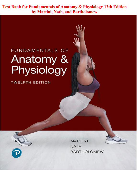
This comprehensive test bank is built for the 12th Edition of Fundamentals of Anatomy & Physiology by Martini, Nath, and Bartholomew. It is one of the most comprehensive, most scientifically rigorous, and most widely used anatomy and physiology textbooks in pre-health science and nursing education — and this test bank helps you master every chapter with the focused, mechanistically grounded, clinically applied practice that A&P mastery demands.
Whether you are completing a pre-health science prerequisite, preparing for a course exam, building toward the NCLEX, or establishing the scientific foundation for any healthcare career, this resource delivers the systematic, body-system-by-body-system, clinical-application-connected practice that the 12th Edition of Martini, Nath, and Bartholomew demands.
What’s Inside?
- Hundreds of practice questions covering every chapter
- Multiple-choice, true/false, matching, and clinical application questions
- Complete answer keys with thorough, mechanistically grounded rationales
- Questions covering anatomical structures, physiological mechanisms, molecular biology applications, and clinical disease connections
- Content fully aligned with the 12th Edition’s updated chapter organization, expanded clinical content, and current anatomical terminology
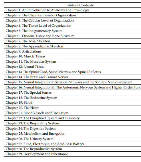
Table of Contents
Unit 1: Levels of Organization
- An Introduction to Anatomy and Physiology
- The Chemical Level of Organization
- The Cellular Level of Organization
- The Tissue Level of Organization
Unit 2: Support and Movement
- The Integumentary System
- Bones and Bone Structure
- The Axial Skeleton
- The Appendicular Skeleton
- Joints
- Muscle Tissue
- The Muscular System
Unit 3: Control and Regulation
- Nervous Tissue
- The Spinal Cord, Spinal Nerves and Spinal Reflexes
- The Brain and Cranial Nerves
- Sensory Pathways and the Somatic Nervous System
- The Autonomic Nervous System and Higher Order Functions
- The Special Senses
- The Endocrine System
Unit 4: Fluids and Transport
- Blood
- The Heart
- Blood Vessels and Circulation
- The Lymphatic System and Immunity
Unit 5: Environmental Exchange
- The Respiratory System
- The Digestive System
- Metabolism, Nutrition and Energetics
- The Urinary System
- Fluid, Electrolyte and Acid-Base Balance
Unit 6: Continuity of Life
- The Reproductive System
- Development and Inheritance
Who Is This Test Bank For?
This resource is perfect for:
- Pre-nursing students completing anatomy and physiology prerequisites using Martini, Nath, and Bartholomew
- Pre-medical, pre-dental, pre-pharmacy, and pre-physical therapy students in rigorous A&P courses
- Undergraduate nursing students reviewing foundational anatomy and physiology concepts
- Allied health students in respiratory therapy, radiography, and surgical technology programs
- Science instructors building course exams and unit assessments for the 12th Edition
- Healthcare professionals seeking a comprehensive foundational science refresher
- Graduate students reviewing anatomical and physiological foundations for advanced coursework
- Students whose programs have adopted the updated 12th Edition
Topics Covered Include:
- An introduction to anatomy and physiology — organization, homeostasis, and anatomical terminology
- The chemical level of organization — atoms, molecules, and biochemistry
- The cellular level of organization — cell structure, function, and cellular communication
- The cellular level — membrane transport, gene expression, and cell division
- The tissue level of organization — epithelial, connective, muscular, and nervous tissues
- The integumentary system — skin structure, functions, and clinical conditions
- Osseous tissue and bone structure — bone physiology and calcium homeostasis
- The skeletal system — the axial skeleton
- The skeletal system — the appendicular skeleton
- Joints — classification, structure, and movements
- Muscle tissue — structure, contraction mechanism, and fiber types
- The muscular system — major muscle groups, origins, insertions, and actions
- Neural tissue — neuron structure, membrane potentials, and synaptic transmission
- The central nervous system — the brain and spinal cord
- The peripheral nervous system — sensory receptors and spinal nerves
- The autonomic nervous system — sympathetic and parasympathetic divisions
- Sensory and motor tracts and the spinal cord
- The special senses — olfaction, gustation, vision, hearing, and equilibrium
- The endocrine system — hormones, glands, and regulatory mechanisms
- Blood — composition, blood types, clotting, and hematopoiesis
- The heart — cardiac anatomy, physiology, and the cardiac cycle
- Blood vessels and circulation — vascular anatomy and hemodynamics
- The lymphatic system and immunity — innate and adaptive immune responses
- The respiratory system — pulmonary anatomy, ventilation, and gas exchange
- The digestive system — organs, digestive processes, and nutrient absorption
- Metabolism and energetics — cellular metabolism and metabolic regulation
- The urinary system — kidney anatomy, nephron function, and urine formation
- Fluid, electrolyte, and acid-base balance
- The reproductive systems — male and female anatomy, hormonal regulation, and gametogenesis
- Development and inheritance — fertilization, embryology, and genetics
Why This Test Bank Delivers Results
Martini, Nath, and Bartholomew’s Fundamentals of Anatomy & Physiology has earned its standing as one of the most authoritative pre-health science A&P textbooks through a consistent commitment to scientific accuracy, mechanistic depth, and clinical relevance that few competing textbooks match.
The 12th Edition raises that standard further — with updated molecular and cellular biology content reflecting the most current biomedical research, expanded clinical application throughout every chapter, refreshed disease examples incorporating the most current pathophysiological understanding, enhanced coverage of homeostatic mechanisms at the molecular and cellular level, and the most current anatomical terminology consistent with international anatomical naming conventions.
This test bank is built to develop the mechanistic, integrated understanding that Martini, Nath, and Bartholomew demand.
Every question requires you to apply anatomical knowledge and physiological reasoning to clinical scenarios — not simply recall isolated facts from a chapter summary. You will trace the cascade of events that occurs when serum calcium falls below normal — identifying each step from parathyroid hormone secretion through osteoclast activation to renal calcium reabsorption — and predict the consequences of disrupting any step in that cascade. You will follow a molecule of oxygen from atmospheric air through the respiratory passages, across the alveolar-capillary membrane, into hemoglobin, through the cardiovascular circuit, and into a mitochondrion — and explain the clinical consequences of impairment at any point along that pathway. You will analyze a complete blood count and apply your knowledge of hematopoiesis and blood cell function to identify what each abnormal value suggests about the patient’s underlying pathology.
This is the level of anatomical and physiological mastery that Martini, Nath, and Bartholomew produce in their students — and that healthcare educators consistently recognize as the preparation standard for rigorous health science programs. And this test bank builds it — question by question, mechanism by mechanism, clinical connection by clinical connection.
Detailed rationales explain the anatomical structures, physiological mechanisms, molecular biology, and clinical disease significance behind every correct answer — reflecting the same scientific depth and clinical integration that the 12th Edition establishes throughout. Questions are organized chapter by chapter for structured, systematic study.
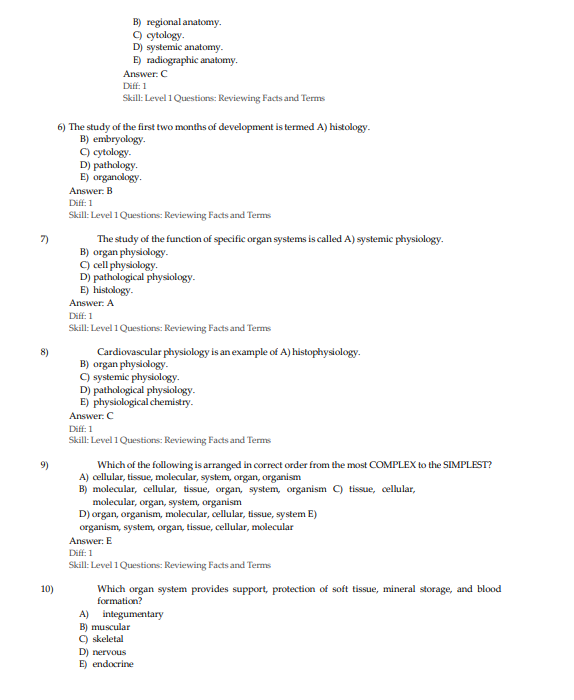
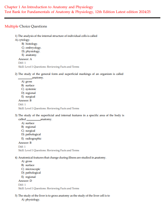
Sample Questions
Question 1
A student is studying the sliding filament theory of muscle contraction. A patient with myasthenia gravis has autoantibodies that destroy nicotinic acetylcholine receptors at the neuromuscular junction. Tracing the mechanism of muscle contraction from nerve impulse to cross-bridge cycling, at which specific step does the pathological process in myasthenia gravis interrupt normal muscle contraction?
- A) The action potential propagating along the motor neuron’s axon is blocked, preventing depolarization from reaching the axon terminal
- B) Acetylcholine release from synaptic vesicles at the axon terminal is inhibited, preventing neurotransmitter from reaching the motor end plate
- C) Acetylcholine cannot bind to its postsynaptic receptors because they have been destroyed by autoantibodies — preventing depolarization of the motor end plate, generation of a muscle action potential, and initiation of the excitation-contraction coupling cascade
- D) Calcium release from the sarcoplasmic reticulum is blocked, preventing troponin-tropomyosin complex displacement and active site exposure on actin
Correct Answer: C
Rationale: This question requires tracing the complete neuromuscular transmission sequence and identifying the specific disruption produced by myasthenia gravis pathophysiology. Normal neuromuscular transmission proceeds through the following sequence — a nerve action potential reaches the axon terminal, triggering voltage-gated calcium channel opening and calcium influx into the terminal, which drives synaptic vesicle fusion with the presynaptic membrane and exocytosis of acetylcholine into the neuromuscular junction. Acetylcholine diffuses across the junction and binds to nicotinic acetylcholine receptors on the postsynaptic motor end plate, opening ligand-gated sodium channels, generating a motor end plate potential, and initiating a muscle action potential that propagates along the sarcolemma and down the T-tubules. T-tubule depolarization opens ryanodine receptor calcium channels on the sarcoplasmic reticulum, releasing calcium into the sarcoplasm. Calcium binds to troponin C on the troponin-tropomyosin complex, causing tropomyosin to shift and expose myosin-binding sites on actin, allowing cross-bridge cycling and force generation. In myasthenia gravis, IgG autoantibodies bind to and destroy nicotinic acetylcholine receptors at the motor end plate. Despite normal acetylcholine release, the neurotransmitter cannot bind to sufficient functional receptors to generate adequate motor end plate depolarization — the transmission fails at the receptor level. This explains the characteristic fatigable muscle weakness of myasthenia gravis — with activity, available receptors are progressively occupied and degraded, worsening the transmission failure. The axon potential propagation and acetylcholine release are normal — the block is postsynaptic.
Question 2
A physiology student is studying the renin-angiotensin-aldosterone system and its role in blood pressure and fluid balance regulation. A patient with bilateral renal artery stenosis develops severe hypertension and secondary hyperaldosteronism. Which complete physiological cascade explains how reduced renal perfusion from the stenoses produces systemic hypertension and hyperaldosteronism?
- A) Reduced renal perfusion directly stimulates the adrenal cortex to secrete aldosterone, which causes water retention and hypertension without renin involvement
- B) Reduced renal perfusion is detected by the juxtaglomerular apparatus, which secretes renin — renin cleaves angiotensinogen to angiotensin I — ACE in the pulmonary endothelium converts angiotensin I to angiotensin II — angiotensin II causes direct vasoconstriction and stimulates adrenal cortex aldosterone secretion — aldosterone drives renal sodium and water reabsorption — the combined vasoconstriction and volume expansion produce hypertension
- C) Reduced renal perfusion causes the kidney to directly secrete angiotensin II without requiring renin activation, bypassing the normal cascade
- D) Renal artery stenosis reduces urine output, causing fluid accumulation that directly elevates blood pressure without hormonal mediation
Correct Answer: B
Rationale: This question requires tracing the complete RAAS cascade from the triggering stimulus through every intermediary step to the final hemodynamic consequence — the level of mechanistic understanding that Martini, Nath, and Bartholomew consistently demand. The juxtaglomerular cells of the kidney detect decreased stretch in the afferent arteriole wall — reflecting reduced perfusion pressure from the stenotic renal artery — as well as decreased sodium delivery to the macula densa, as a secondary consequence of reduced filtration. These stimuli trigger renin secretion from the JGA into the circulation. Renin is a protease enzyme that cleaves the hepatically produced plasma protein angiotensinogen, releasing the decapeptide angiotensin I. Angiotensin I has minimal biological activity until it reaches the pulmonary vascular endothelium, where angiotensin-converting enzyme cleaves two amino acids to produce the highly active octapeptide angiotensin II. Angiotensin II exerts its hypertensive effects through two primary mechanisms — direct, potent vasoconstriction of arterioles throughout the systemic circulation, raising peripheral vascular resistance and blood pressure; and stimulation of the adrenal cortex zona glomerulosa to synthesize and secrete aldosterone. Aldosterone acts on the principal cells of the renal collecting duct to upregulate sodium-potassium ATPase pumps and apical sodium channels, increasing sodium reabsorption with osmotic water following, expanding extracellular fluid volume. The combination of increased peripheral resistance and increased circulating volume drives the severe hypertension observed in bilateral renal artery stenosis — explaining why ACE inhibitors are particularly effective in this condition but require careful monitoring for renal function deterioration.
Question 3
A student is studying the physiology of the immune system. A patient undergoes a solid organ transplant and requires immunosuppressive therapy with tacrolimus, which inhibits T-cell activation by blocking calcineurin — a phosphatase required for interleukin-2 gene transcription. Which specific component of the adaptive immune response does tacrolimus most directly target, and what is the physiological consequence of this inhibition?
- A) Tacrolimus inhibits B-cell antibody production by blocking immunoglobulin heavy-chain gene rearrangement in activated B lymphocytes
- B) Tacrolimus inhibits T-cell receptor signaling by preventing the activation of calcineurin — which normally dephosphorylates NFAT and allows it to enter the nucleus and drive IL-2 transcription — reducing T-cell proliferation, clonal expansion, and both cytotoxic T-cell and T-helper cell responses that would otherwise drive transplant rejection
- C) Tacrolimus eliminates natural killer cell activity by blocking perforin and granzyme release from NK cell granules
- D) Tacrolimus suppresses the complement system by blocking C3 convertase formation and preventing the membrane attack complex from assembling
Correct Answer: B
Rationale: This question integrates the molecular mechanisms of T-cell activation with the pharmacology of immunosuppression — a clinical application of adaptive immunity physiology that Martini, Nath, and Bartholomew specifically address in their coverage of the immune system and its clinical modulation. When a T-cell receptor recognizes a foreign antigen presented by an MHC molecule, a complex intracellular signaling cascade is initiated — including activation of calcineurin, a calcium-dependent phosphatase. Calcineurin dephosphorylates the Nuclear Factor of Activated T-cells transcription factor, allowing NFAT to translocate into the nucleus and drive transcription of interleukin-2 and other cytokine genes critical for T-cell activation, proliferation, and differentiation. IL-2 functions as the primary T-cell growth factor — it drives clonal expansion of both cytotoxic CD8+ T-cells and CD4+ T-helper cells that would otherwise orchestrate cellular rejection of the transplanted organ. Tacrolimus binds to the cytoplasmic protein FKBP-12, and the tacrolimus-FKBP12 complex specifically inhibits calcineurin’s phosphatase activity. Without calcineurin activity, NFAT remains phosphorylated in the cytoplasm, cannot translocate to the nucleus, and IL-2 transcription is dramatically reduced. The result is profound suppression of T-cell activation and proliferation — preventing the cytotoxic and helper T-cell responses that constitute the primary cellular immune mechanism of organ rejection. Tacrolimus does not directly affect B-cell immunoglobulin gene rearrangement, NK cell perforin release, or complement system activation — its primary mechanism is specifically T-cell calcineurin inhibition.
Question 4
A student is reviewing the physiology of the renal tubule and the mechanisms of acid-base balance. A patient develops metabolic alkalosis following three days of nasogastric suction with continuous removal of gastric hydrochloric acid. Tracing the renal compensatory response, which specific tubular mechanism most directly works to restore blood pH toward normal in this patient?
- A) The kidneys increase bicarbonate secretion into the tubular fluid through the apical chloride-bicarbonate exchanger of the proximal tubule, reducing plasma bicarbonate concentration to normalize pH
- B) The kidneys increase H⁺ ion reabsorption from the tubular fluid back into the peritubular capillaries, reducing tubular acidification to correct the alkalosis
- C) The kidneys reduce bicarbonate reabsorption in the proximal tubule and increase bicarbonate excretion in the collecting duct — reducing plasma bicarbonate concentration — while decreasing H⁺ secretion — reducing tubular acidification — to allow pH to normalize
- D) The kidneys activate the renin-angiotensin-aldosterone system to increase sodium reabsorption, which indirectly reduces blood pH through sodium-hydrogen exchange
Correct Answer: C
Rationale: Understanding renal compensation for metabolic alkalosis requires mechanistic knowledge of how the kidney regulates plasma bicarbonate through tubular reabsorption, secretion, and generation. In normal acid-base physiology, the kidney’s default tendency is to reabsorb bicarbonate and secrete hydrogen ions — this conserves bicarbonate and eliminates acid. When metabolic alkalosis develops — from continuous HCl loss in this scenario — the plasma bicarbonate concentration rises above normal as the acid-base equation shifts. The renal compensatory response reverses the kidney’s default tendency. In the proximal tubule, sodium-hydrogen exchangers — normally responsible for H⁺ secretion and indirect bicarbonate reabsorption — reduce their activity. In the collecting duct, alpha-intercalated cells — which normally secrete H⁺ and reabsorb bicarbonate — reduce their activity, while beta-intercalated cells — which normally secrete bicarbonate — increase bicarbonate secretion into the tubular fluid. The net effect is reduced bicarbonate reabsorption and increased bicarbonate excretion in the urine — causing plasma bicarbonate to fall back toward normal and blood pH to normalize. The kidneys do not reabsorb H⁺ from tubular fluid — hydrogen ion secretion is unidirectional from blood into tubular lumen. RAAS activation would worsen metabolic alkalosis through enhanced sodium-hydrogen exchange — not correct it. The renal compensation for metabolic alkalosis is producing alkaline urine with high bicarbonate content.
Question 5
A student is studying the endocrine system and the hormonal regulation of blood glucose. A patient with type 1 diabetes mellitus experiences absolute insulin deficiency during a period of insulin pump failure. Tracing the metabolic consequences of absolute insulin deficiency, which cascade of events most completely explains the development of diabetic ketoacidosis in this patient?
- A) Absolute insulin deficiency causes the pancreas to overproduce glucagon, which directly stimulates insulin secretion from residual beta cells — creating a compensatory hyperinsulinemic state that paradoxically drives DKA
- B) Absolute insulin deficiency removes the inhibitory signal on glucagon secretion from alpha cells — glucagon drives hepatic glycogenolysis and gluconeogenesis causing hyperglycemia — simultaneously, insulin deficiency prevents glucose uptake by peripheral tissues — counter-regulatory hormone-driven lipolysis floods the liver with free fatty acids which are converted to ketone bodies — ketone accumulation causes high anion gap metabolic acidosis while osmotic diuresis from hyperglycemia causes dehydration
- C) Absolute insulin deficiency causes the kidneys to produce excess bicarbonate in compensation for rising blood glucose, generating a metabolic alkalosis rather than acidosis
- D) Absolute insulin deficiency has no effect on lipid metabolism — DKA results exclusively from the osmotic consequences of hyperglycemia without ketogenesis
Correct Answer: B
Rationale: This question requires integrating multiple physiological systems — endocrine regulation, carbohydrate metabolism, lipid metabolism, acid-base physiology, and renal fluid handling — to trace the complete DKA pathophysiological cascade. In type 1 diabetes, the autoimmune destruction of pancreatic beta cells eliminates endogenous insulin production. During pump failure, exogenous insulin is also absent. Without insulin, glucagon — normally suppressed by insulin — acts unopposed from pancreatic alpha cells. Glucagon drives hepatic glycogenolysis — breaking down glycogen stores — and gluconeogenesis — synthesizing glucose from amino acids and glycerol — dramatically elevating blood glucose. Simultaneously, without insulin’s anabolic signal, peripheral tissues — skeletal muscle and adipose — cannot take up glucose through GLUT4 transporters, leaving cells in a state of intracellular starvation despite extreme extracellular hyperglycemia. This intracellular starvation, combined with cortisol and catecholamine counter-regulatory hormone release, drives lipolysis — hormone-sensitive lipase breaks down triglycerides, releasing free fatty acids into the circulation. The liver takes up these free fatty acids and, in the absence of insulin which would promote fatty acid storage, converts them through beta-oxidation into acetyl-CoA — which enters the ketogenic pathway generating acetoacetate and beta-hydroxybutyrate. These ketone bodies accumulate in the blood, dissociate into H⁺ and their anion, consuming bicarbonate buffers and producing high anion gap metabolic acidosis. Glucose above the renal threshold — approximately 180 mg/dL — spills into the urine, creating an osmotic diuresis that causes profound polyuria, dehydration, and electrolyte loss. The complete DKA picture — hyperglycemia, ketoacidosis, and dehydration — emerges from this integrated metabolic cascade.
Frequently Asked Questions (FAQs)
What edition does this test bank cover?
This test bank is written specifically for the 12th Edition of Fundamentals of Anatomy & Physiology by Martini, Nath, and Bartholomew. All questions are fully aligned with the current edition’s chapter organization, updated molecular and cellular biology content, expanded clinical application, and current anatomical terminology standards.
How are the questions organized?
Questions are arranged chapter by chapter — body system by body system — allowing you to study systematically from the chemical level of organization through development and inheritance, or focus specifically on the systems where your examination performance needs the most improvement.
How does this test bank differ from test banks for other A&P textbooks?
This test bank is built to match the scientific depth and mechanistic rigor of Martini, Nath, and Bartholomew’s 12th Edition — one of the most comprehensive A&P textbooks in pre-health science education. Questions go beyond anatomical memorization to require mechanistic physiological reasoning, molecular biology application, and clinical disease connection — reflecting the depth the 12th Edition demands.
Is this test bank useful for nursing school prerequisite preparation?
Absolutely. Fundamentals of Anatomy & Physiology by Martini, Nath, and Bartholomew is one of the most commonly required A&P textbooks for nursing and health science prerequisites. This test bank is directly aligned with the 12th Edition and provides the scientific depth and clinical application that rigorous healthcare prerequisite courses require.
Is this test bank appropriate for pre-medical and allied health students?
Yes. This textbook and test bank are used across pre-health science disciplines — pre-nursing, pre-medical, pre-dental, pre-pharmacy, pre-physical therapy, respiratory therapy, and more. Any student using the 12th Edition of Martini, Nath, and Bartholomew will find this test bank directly relevant to their coursework.
How quickly can I access the test bank after purchase?
Immediately. As soon as your purchase is complete, you receive instant digital access with no waiting period. Study on your own schedule, at your own pace, from any device.
Are the rationales written at the mechanistic depth that Martini, Nath, and Bartholomew demand?
Yes. Every rationale explains the anatomical structure, physiological mechanism, molecular biology, and clinical significance behind the correct answer at the level of depth the 12th Edition establishes — building the mechanistic understanding that distinguishes students who truly master A&P from students who merely memorize it.
Can instructors use this test bank for course assessments?
Absolutely. The chapter-by-chapter organization, mechanistic question construction, molecular biology integration, clinical application emphasis, and scientific rigor make this an outstanding resource for faculty building unit exams, quizzes, and comprehensive course assessments for anatomy and physiology courses using the 12th Edition of Martini, Nath, and Bartholomew.



Caleb Martins –
Perfect
Lucas Gabriel –
Excellent test bank for efficient exam preps
Mary Kesh –
A solid study guide
Helen Christie –
Much better than I expected. Thanx
Dorcas M. –
This guide helped me nail my midterms and finals
Melania Angel –
I like it
Kamara Reid –
Comprehensively covers all important topics
Rosemary R –
The resource made my work a lot easier
Liam Mbogo –
A lot of questions from this test bank featured in my final exam
Mary Lincoln –
Helped me stay consistent with nursing exam preparation.
Morgan Fraiser –
Very helpful for NCLEX-style practice.
Peterson S. –
Excellent resource for exam prep
Leo Fernandez –
Excellent way to practice before exams.
Jarrod Bent –
Boosted my A&P exam confidence
Cayte N. –
Simple explanations made reviewing answers much easier.